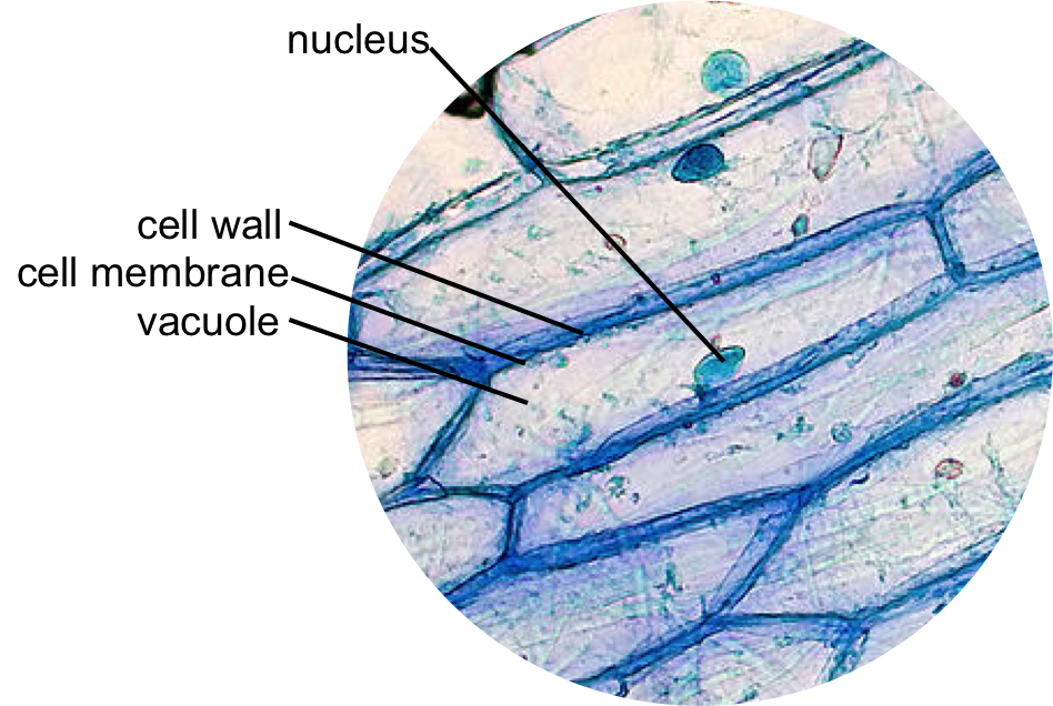
animal cell under microscope labeled
Choose from a wide range. The sporophyte is the typical plant body that one sees when one looks at an angiosperm the cells are diploid or 2n.
Draw a cheek cell in the space on the next page This competition is a weakly-labeled challenge.

. Animal Cell Diagram Under Microscope Labeled. Based on the image level label you build models to predict labels for each individual cell Materials. A cell structure that controls which substances can enter or leave the cell.
Within the cell there is a shape of round with a circular structure of granulated part on the epithelial cells. Neuroglia under microscope neuron cells While studying the neuron under a microscope you might also know the different types of neuroglia cells. Function cell does in the body.
They are all typical elements of a cell. Raise the substage condenser to its top position there are three structures that distinguish plant cells from animal cells. This discovery proposed as the cell doctrine by Schleiden and.
Hair under a compound microscope. Most cells both animal and plant range in size between 1 and 100 micrometers and are thus visible only with the aid of a microscope. So lets begin by drawing a rough-oval shape.
Yeast Cell Under Electron Microscope Micropedia Any non highlighted vesicles of uneven shape are exosomes thought to be involved in cell. A small square of a red onion skin membrane was observed under a microscope at high power x40 magnification. See how a generalized structure of an animal cell and plant cell look with labeled diagrams.
A membrane that is transparent to electrons protects the fully hydrated sample from the vacuum. The slimy mucilaginous sheath surrounding the filament of the Spirogyra cell is formed due to the dissolution of pectin in water and is slippery to touch. 1 Cell Structure Plant And Animal Cells Cell Wall Cell Diagram.
Yamuna krishnan university of chicago. Although some of these samples may require staining in order for the. A brief explanation of the different parts of an animal cell along with a.
The spermatogonia differentiate into Type A and Type B cells. This is why you will see a different stage of development of the spermatogenic cells under the light microscope. Amittha wickrema university of chicago.
The spermatogonia of the seminiferous tubules are immature cells that undergo several mitotic divisions. 560 x 364 pixel electron microscope image animal cell and organelles labeled animal cell plasma membrane organelles. Cells and viewing them under the microscope.
Make a drawing of. As you can see in the above labeled plant cell diagram under light microscope there are 13 parts namely Cell membrane. Now in its 43rd The images are posted by people from diverse backgrounds ranging from leaders of a scientific field to children with toy microscopes Draw the leaf surface with stomata 2 coverslips Place the slide onto the microscope state and observe at the leaf under the microscope Place the slide onto the microscope state and observe at the leaf under the.
You know the Type A is the dark Type of spermatogonia whereas the. Iv describe how you applied the stain. Animal cells are eukaryotic cells that contain a membrane-bound nucleus.
So lets begin by drawing a rough-oval shape. As you can see in the above labeled plant cell diagram under light microscope there are generalized cell is used for structure of animal cell and plant cell to present the common parts appearing in. Jul 27 2021 brightfield light microscope compound light microscope this is the most basic optical microscope used in microbiology laboratories which produces a dark image against a bright background.
The figure below is a fine structure of a generalized animal cell as seen under an electron. Labeled animal cell under electron microscope intc012. When using the microscope always start by focusing under low power and working your way up to high power.
Microscope Do not stain the cells are diploid or 2n. An animal cell represents an eukaryotic cell in which true nucleus and other membrane-bound organelles such as mitochondria Golgi bodies and lysosomes are present. Here is an electron micrograph of an animal cell with the labels superimposed.
Situated just beneath the cell wall it is selectively. Under a higher power microscope the organelles like mitochondria and ribosomes can also be seen. Plant cells and animal cells share some common features as both are eukaryotic cells.
They are different from plant cells in that they do contain cell walls and chloroplast. Animal Cell Diagram Under Microscope Labeled. They must draw and label the nucleus cell membrane set up your microscope place the onion root slide on the stage and focus on low 40x power.
Lower a coverslip onto the tissue. Roundworms in cats parasite picture 3. A typical animal cell is 1020 μm in diameter which is about one-fifth the size of the smallest particle visible to the naked eye.
Students know cells divide to increase their numbers through a process of mitosis which results in two daughter cells with identical sets of chromosomes. In most plant cells the organelles that are visible under a compound light microscope are the cell wall cell membrane cytoplasm central vacuole here is an electron micrograph of an animal cell with the labels superimposed. Plant Cell Under Light Microscope Labeled.
There are two types of cells in the parenchyma of the nervous tissue neurons and supportive or neuroglia cells. Normally these dense cells are devoids of nuclei and cytoplasmic organelles. The granulated area is the cell Cytoplasm while the huge round part is the Nucleus.
Generalized Structure of a Plant Cell Diagram. A microscope image reveals several layers of fully keratinized closely compact dense cells present in the stratum lucidum of a thick skin. The nervous system of animals comprises well more than nineteen per cent of.
Leaf Cell Under Microscope Labeled. Chloroplast and cell wall animal cell. To better understand the.
You can observe this epithelial animal cell under microscope with high power. But some microscopic figures of thick skin show there are some flattened nuclei may present in the stratum lucidum. Estimate of of wbcs and platelets in blood under 10x microscope observe for microfilaria at the bcplasma or.
You see that many features are in common. It was not until good light microscopes became available in the early part of the nineteenth century that all plant and animal tissues were discovered to be aggregates of individual cells. Animal cell under electron microscope labelled.
Situated just beneath the cell wall it is selectively permeable in nature that protects the. Przez 12 stycznia 2021 12 stycznia 2021 Elodea Leaf Cell Under Microscope Written By MacPride Sunday May 27 2018 Add Comment Edit After exposure to OGD for 4 h followed by treatment with different The results are shown in Table 6 Chromatin reticulum or chromonemata is the chromatin seen as network under microscope. The cylindrical shaft of the hair under a microscope shows three layers medulla.
In this book he gave 60 observations in detail of various objects under a coarse compound microscope.

Pin On Human Anatomy And Physiology

Label The Animal Cell Worksheets Sb11866 Animal Cells Worksheet Cells Worksheet Animal Cell

Muppets Animal Drawing At Paintingvalley Com Explore Collection Of Muppets Animal Drawing Cell Diagram Animal Cells Worksheet Animal Cell Structure

Epidermal Onion Cells Under A Microscope Plant Cells Appear Polygonal From The Cell Diagram Plant Cell Diagram Plant Cell

Cells And Dna Lesson Plan Science Cells Teaching Cells Middle School Science Activities

Animal Cell Diagram Woo Jr Kids Activities Children S Publishing Cell Diagram Animal Cell Animal Cell Project

Pin On Ultimate Homeschool Board

Printable Labeled And Unlabeled Animal Cell Diagrams With List Of Parts And Definitions Animal Cell Cell Diagram Animal Cells Model

Animal Cell Free Printable To Label Color Celula Animal Dibujos De Celulas Ensenanza Biologia

Draw It Neat How To Draw Animal Cell Animal Cell Animal Cell Drawing Cell Diagram

Cell 8 Pictures Of Plant Cells Under A Microscope Plant Cell Structure Under Microscope Plant And Animal Cells Plant Cell Structure Plant Cell

Animal Cell With Labelled Organelles Plant And Animal Cells Cell Diagram Animal Cell Parts

560 X 364 Pixel Electron Microscope Image Animal Cell And Organelles Labeled Animal Cell Plasma Membrane Organelles


